Netter’s Atlas of Human Anatomy PDF: A Comprehensive Overview
Netter’s Atlas stands as a cornerstone resource for anatomy education, offering detailed visual guidance.
Numerous editions, including the sixth and eighth, are available in PDF format through platforms like the Internet Archive.
Medical students frequently utilize this atlas for its clear illustrations and regional organization, aiding in the comprehension of complex anatomical structures.
Netter’s Atlas of Human Anatomy has long been recognized as a premier anatomical resource for medical, dental, and allied health students, as well as practicing professionals. Its enduring popularity stems from a unique combination of artistic clarity and anatomical accuracy. The atlas doesn’t simply present anatomical facts; it visually communicates the spatial relationships of structures, fostering a deeper understanding of the human body.
Initially conceived by Dr. Frank H. Netter, the atlas began as a personal project to improve his own understanding and teaching of anatomy. This dedication to clarity and precision is evident in every illustration. Today, the atlas continues to evolve with each new edition, incorporating advancements in medical imaging and anatomical knowledge. Access to Netter’s Atlas is readily available, with PDF versions frequently sought for convenient study and reference, often found through resources like the Internet Archive.
The atlas serves as an invaluable companion to anatomy coursework, providing a visual guide that complements textbook descriptions and laboratory dissections.
Historical Context and Frank H. Netter
Frank H. Netter (1906-1991), a physician and accomplished medical illustrator, laid the foundation for what would become the gold standard in anatomical atlases. Dissatisfied with the limited visual resources available during his medical training, Netter began creating his own illustrations to better understand and teach anatomy. These initial sketches, born from a need for clarity, evolved into a comprehensive collection that ultimately formed the basis of Netter’s Atlas.
The first edition of the atlas was published in 1983, quickly gaining recognition for its exceptional detail and artistic quality. Netter’s commitment to accuracy and his ability to visually represent complex anatomical relationships set his work apart. His legacy continues through subsequent editions, meticulously updated by a team of expert anatomists. The availability of Netter’s Atlas in PDF format has broadened its accessibility, allowing students and professionals worldwide to benefit from Netter’s pioneering work.
Significance for Medical Students
Netter’s Atlas of Human Anatomy holds immense significance for medical students navigating the complexities of gross anatomy. Traditional learning often relies heavily on memorization, but Netter’s provides a crucial visual complement, transforming abstract concepts into tangible, understandable forms. The atlas’s detailed illustrations facilitate spatial reasoning, enabling students to visualize structures in three dimensions and grasp their relationships within the body.
The regional approach employed by Netter’s mirrors the way anatomy is often taught in medical curricula, making it a seamless study companion. Access to the PDF version offers unparalleled convenience, allowing students to study anytime, anywhere. Furthermore, the atlas aids in clinical correlation, bridging the gap between anatomical knowledge and real-world medical practice. Mastering anatomy with Netter’s builds a strong foundation for future success in medical studies and beyond.
Key Features of the Atlas
Netter’s Atlas is renowned for its beautifully illustrated, accurate depictions of human anatomy. Its regional organization and inclusion of clinical correlations enhance understanding and learning.
Illustrative Style and Accuracy
Netter’s Atlas of Human Anatomy distinguishes itself through a unique illustrative style, meticulously crafted by Frank H. Netter himself. These aren’t simply diagrams; they are artistic renderings that prioritize clarity and anatomical precision. The illustrations emphasize a realistic representation of structures, utilizing color coding and consistent viewpoints to facilitate learning.
Accuracy is paramount. Each illustration undergoes rigorous review by medical experts to ensure fidelity to anatomical detail. The atlas doesn’t just show anatomy; it aims to present it as it appears in the human body, aiding in spatial understanding. This commitment to both artistry and scientific correctness has made Netter’s the gold standard for generations of medical students and professionals. The detailed nature of the illustrations allows for a deeper comprehension of complex anatomical relationships.
Regional Approach to Anatomy

Netter’s Atlas of Human Anatomy employs a distinctly regional approach to the study of the human body. Rather than dissecting systems in isolation, the atlas presents anatomy organized by body region – head and neck, upper limb, lower limb, and so on. This method mirrors how anatomy is often encountered in clinical practice, fostering a more practical understanding.
Within each region, structures are presented layer by layer, building from superficial to deep. This layered approach allows students to visualize the relationships between different anatomical elements. The atlas systematically covers muscles, nerves, vessels, and bones within each region, providing a comprehensive overview. This organization facilitates spatial reasoning and helps learners build a robust mental map of the human anatomy, enhancing clinical application.
Integration of Clinical Relevance

Netter’s Atlas of Human Anatomy doesn’t present anatomy in a vacuum; it actively integrates clinical relevance throughout its illustrations and accompanying text. While primarily an anatomical resource, the atlas frequently highlights structures pertinent to common clinical scenarios, such as sites for injections, surgical approaches, and potential areas of injury.
This integration helps students understand why anatomical knowledge is crucial for medical practice. The atlas subtly connects anatomical landmarks to clinical presentations, aiding in diagnostic reasoning. For example, nerve pathways are often depicted in relation to potential compression points. This approach transforms the learning process from rote memorization to a more meaningful understanding of how anatomy impacts health and disease, ultimately preparing students for real-world clinical challenges.

Available Editions and Formats
Netter’s Atlas exists in multiple editions – sixth and eighth being prominent – and formats. PDF and eBook accessibility are widespread, offering convenient digital access to this essential resource.
Sixth Edition Details
The Sixth Edition of Netter’s Atlas of Human Anatomy, authored by Frank H. Netter, M.D., remains a widely accessible and valuable resource, particularly through platforms like the Internet Archive. It’s available for free download, borrowing, and streaming, presented as a PDF file alongside text file options for convenient study.
This edition meticulously illustrates the human anatomy, with a specific example showcasing the esophagus in situ. The Internet Archive provides access to this classic edition, preserving its educational value for current and future learners. The catalog record details its organization into sections covering the upper and lower limbs, alongside cross-sectional anatomy.
Notably, the edition is also identified with specific identifiers like urn:oclc:record:763159902 and urn:lcp:atlasofhumananat0000nett_w1j0, facilitating its location and access within the digital archive. Its enduring popularity stems from Netter’s renowned illustrative style and comprehensive coverage.
Eighth Edition Updates
The Eighth Edition of Netter’s Atlas of Human Anatomy, authored by Frank H. Netter, builds upon the foundation of previous editions with updated illustrations and enhanced clinical correlations. Access to this edition in PDF and ePUB formats is readily available, alongside other medical and anatomy texts.
While specific details regarding the precise updates aren’t extensively detailed in the provided snippets, the edition signifies a continuation of Netter’s commitment to anatomical accuracy and pedagogical clarity. It represents a modern iteration of a classic resource, catering to the evolving needs of medical education.
The availability of the eighth edition in digital formats ensures accessibility for students utilizing various devices and learning preferences. It maintains the atlas’s reputation as a vital tool for understanding the complexities of human anatomy, offering a comprehensive and visually engaging learning experience.
PDF and eBook Accessibility
Netter’s Atlas of Human Anatomy is widely accessible in both PDF and eBook formats, catering to diverse learning preferences and technological capabilities. The Internet Archive hosts several editions, including the sixth, offering free download, borrowing, and streaming options. This broad availability significantly benefits medical students and healthcare professionals globally.
The PDF format allows for easy portability and offline access, while eBook versions provide features like searchable text and adjustable font sizes. Accessing the atlas digitally streamlines study processes and enhances convenience.
However, users should be mindful of copyright and legality when downloading PDFs, ensuring they obtain the atlas from reliable sources. The availability of both formats underscores the publisher’s commitment to making this essential resource readily available to the medical community.

Content Organization within the Atlas
Netter’s Atlas employs a systematic regional approach, covering the head, neck, upper limb, and lower limb in dedicated sections. Cross-sectional anatomy is also included.
Each section presents detailed illustrations and accompanying text for comprehensive anatomical study.
Head and Neck Section

Netter’s Atlas dedicates substantial coverage to the head and neck, recognizing its anatomical complexity. This section meticulously details cranial bones, muscles of facial expression, and the intricate structures within the oral and nasal cavities.
Students benefit from clear depictions of the vasculature and innervation of the head and neck, crucial for understanding clinical presentations. The atlas illustrates the hyoid bone, larynx, pharynx, and thyroid gland with exceptional clarity.
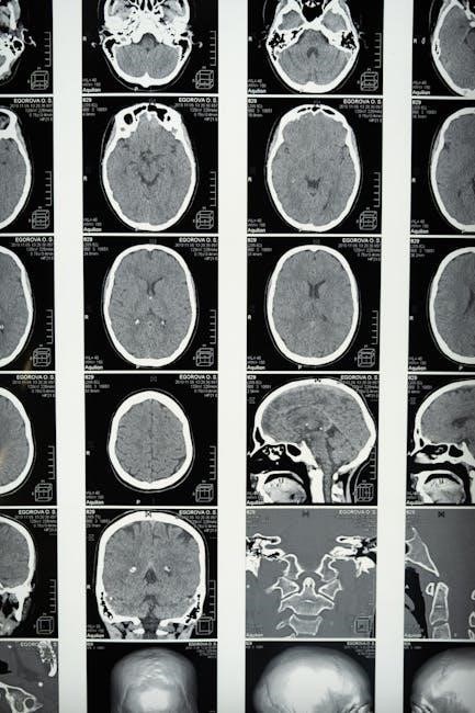
Furthermore, detailed views of the eye, ear, and associated nerves are provided. The section also includes comprehensive coverage of the brain, meninges, and cranial nerves, essential for neurological understanding. The illustrations facilitate spatial reasoning and a deeper grasp of these vital anatomical relationships.
Upper Limb Anatomy
Netter’s Atlas provides a detailed exploration of the upper limb, beginning with the shoulder girdle and progressing distally to the hand. Illustrations clearly depict the muscles, bones, and ligaments of the shoulder, arm, forearm, and wrist.
Particular attention is given to the brachial plexus and associated neurovascular structures, vital for understanding upper limb function and potential clinical issues. The atlas showcases the intricate arrangement of muscles responsible for movements at each joint.
Detailed views of the hand, including palmar and dorsal aspects, highlight the complex network of tendons, ligaments, and intrinsic muscles. Students can readily visualize the carpal tunnel and its contents. The section’s clarity aids in mastering the anatomical basis of upper limb pathologies.
Lower Limb Anatomy
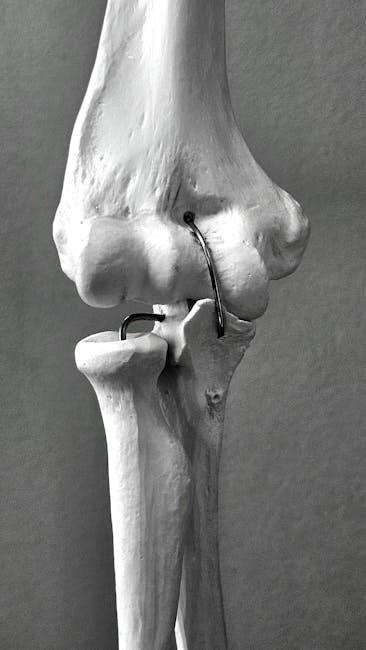
Netter’s Atlas meticulously details the lower limb, starting with the hip and extending through the thigh, leg, and foot. Illustrations showcase the gluteal muscles, femoral triangle, and the complex arrangement of muscles acting on the hip and knee joints.
The atlas provides clear depictions of the popliteal fossa, leg compartments, and the intricate network of arteries, veins, and nerves traversing the lower limb. Special attention is given to the ankle and foot, illustrating the ligaments, tendons, and muscles responsible for stability and movement.
Students benefit from detailed views of the plantar fascia and the intrinsic muscles of the foot. This section facilitates understanding of common lower limb injuries and conditions, solidifying anatomical knowledge for clinical application.

Accessing Netter’s Atlas Online
Netter’s Atlas is accessible online via the Internet Archive, offering free downloads and streaming. However, verifying legality and source reliability is crucial for users.
Internet Archive Availability
The Internet Archive serves as a significant repository for digitized versions of Netter’s Atlas of Human Anatomy, offering both free download and borrowing options. Multiple editions, including the Sixth Edition by Frank H. Netter, M.D., are readily available; Users can access these resources without registration, though creating an account enhances the experience.

Specifically, the archive hosts files in formats like PDF and TXT, allowing for versatile access. The catalog details include identifiers like urn:oclc:record:763159902 and urn:lcp:atlasofhumananat0000nett_w1j0:lcpdf:59c53f96-94a7-4405-a143-c19d8581a0c1, facilitating precise location of desired materials. Furthermore, the archive also contains a French language version, identified as “netter-atlas-d-anatomie-humaine”.
It’s important to note that the availability and specific editions may vary over time, so regular checks are recommended. The Internet Archive’s commitment to open access makes Netter’s Atlas a valuable resource for students and professionals alike.
Free Download Options and Legality
Accessing Netter’s Atlas of Human Anatomy as a free PDF download presents a complex legal landscape. While platforms like the Internet Archive offer access, it’s crucial to understand the terms of use. Borrowing through the Internet Archive is generally considered legal, adhering to controlled digital lending principles.
However, directly downloading PDFs from unofficial sources can infringe on copyright laws. Medical Study Zone highlights the availability of the latest editions for free download, but users should exercise caution and verify the source’s legitimacy.
Purchasing a legitimate copy, whether physical or digital (eBook), ensures compliance with copyright regulations and supports the author and publisher. Always prioritize legal and ethical access to educational materials. Utilizing authorized platforms guarantees access to the most accurate and up-to-date content, avoiding potentially outdated or corrupted files found elsewhere.
Reliable Sources for PDF Downloads
Finding legitimate PDF downloads of Netter’s Atlas requires careful source evaluation. The Internet Archive stands out as a relatively reliable option, offering borrowing access to editions like the sixth, though download availability may vary. Always check the licensing terms before downloading;
Official publisher websites, such as Elsevier, are the most trustworthy sources, though typically require purchase. While some websites advertise “free” downloads, these often lead to compromised files or illegal copies. Caution is paramount when encountering such offers.
University libraries frequently provide students with access to digital versions through their online resources. Exploring these institutional options is a secure and legal pathway. Remember to prioritize sources that respect copyright and offer verified, high-quality content to ensure accurate anatomical study.